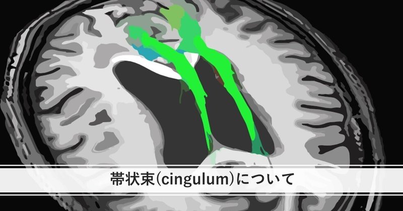
帯状束について

初めまして、くろと申します
臨床4年目、回復期リハビリテーション勤務の作業療法士です。
今回は、帯状束について述べていきたいと思います。
恒例ですが、白質線維とは何かについて話していきます。


ここから帯状束の概説に入ります。

帯状束は前頭葉・頭頂葉・側頭葉とを連絡しています。
さらに、5つのコンポーネントに分かれるとされています。
注意・記憶・感情形成/処理など多くの認知・情動的処理に関与しています。

5) (松田 実 初学者のための神経心理学入門 2022 p292)
6) Wu Y, Sun D, Wang Y, et al: Segmentation of the Cingulum Bundle in the Human Brain: A New Perspective Based on DSI Tractography and Fiber Dissection Study. Front Neuroanat 10: 84, 2016
7) 藤井正純 2019 大脳白質解剖入門 Cadaver Tractography Illustrationで描く,神経科学の温故知新 脳解剖が分かるWEB動画26本付き p31
8) 竹林 崇 2023臨床5年目までに知っておきたい予後予測の考えかた p278
9) Takahashi M, Iwamoto K, Fukatsu H, et al :White matter microstructure of the cingulum and cerebellar peduncle is related to sustained attention and working memory:a diffusion tensor imaging study. Neurosci Lett 477:72—76, 2010
10) Bubb EJ, Metzler—Baddeley C, Aggleton JP:The cingulum bundle:Anatomy, function, and dysfunction. Neurosci Biobe-hav Rev 92: 104—127, 2018
帯状束と連絡する領域のまとめです。
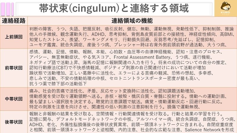

ここから、連絡している領域について説明していきます。
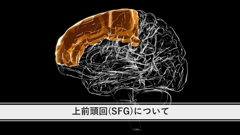
上前頭回の症状は多数あり、主に脱抑制など、高次脳機能障害が生じます。
ADHDとの関連では、親からみた対象者の評価と関連し、右SFGが薄くなっていました。これが不注意や他動に繋がるとも考えられます。また、その結果薄くなったとも考えられます。
思考の抑制と有意な正の相関があり抑制的な機能もありそうです。
左島皮質と右SFGの接続性の増加は精神運動覚醒テストの低下と正の相関がみられます。

18) Hai T, Swansburg R, Kahl CK, Frank H, Stone K, Lemay JF, MacMaster FP. Right Superior Frontal Gyrus Cortical Thickness in Pediatric ADHD. J Atten Disord. 2022 Dec;26(14):1895-1906. doi: 10.1177/10870547221110918. Epub 2022 Jul 11. PMID: 35815438; PMCID: PMC9605998.
19) Lu F, Yang W, Wei D, Sun J, Zhang Q, Qiu J. Superior frontal gyrus and middle temporal gyrus connectivity mediates the relationship between neuroticism and thought suppression. Brain Imaging Behav. 2022;16(3):1400-1409. doi:10.1007/s11682-021-00599-1
20) Wang Y, Yang X, Xiao L, et al. Altered anterior insula-superior frontal gyrus functional connectivity is correlated with cognitive impairment following total sleep deprivation. Biochem Biophys Res Commun. 2022;624:47-52. doi:10.1016/j.bbrc.2022.07.078
21) Wang S, Zhao Y, Zhang L, Wang X, Wang X, Cheng B, Luo K, Gong Q. Stress and the brain: Perceived stress mediates the impact of the superior frontal gyrus spontaneous activity on depressive symptoms in late adolescence. Hum Brain Mapp. 2019 Dec 1;40(17):4982-4993. doi: 10.1002/hbm.24752. Epub 2019 Aug 9. PMID: 31397949; PMCID: PMC6865488.
イライラを抱える若者は健常者と比較して右上前頭回が萎縮し
イライラと負の相関がみられる。
上前頭回は羨望と関連するが、右縁上回・楔前部との接続が大きいほど羨望が少なくなることが示されています。
楔前部がデフォルトモードネットワーク内において空想、視覚イメージなどに関与していることから、羨望の光景をイメージで表現しているかもしれません。その場合大きな接続性によって抑制することで羨望が減少することが考えられます。

23) McDonald B, Becker K, Meshi D, Heekeren HR, von Scheve C. Individual differences in envy experienced through perspective-taking involves functional connectivity of the superior frontal gyrus. Cogn Affect Behav Neurosci. 2020 Aug;20(4):783-797. doi: 10.3758/s13415-020-00802-8. PMID: 32557135; PMCID: PMC7395029.
24) Chen L, Wang Y, Niu C, Zhong S, Hu H, Chen P, Zhang S, Chen G, Deng F, Lai S, Wang J, Huang L, Huang R. Common and distinct abnormal frontal-limbic system structural and functional patterns in patients with major depression and bipolar disorder. Neuroimage Clin. 2018 Jul 6;20:42-50. doi: 10.1016/j.nicl.2018.07.002. PMID: 30069426; PMCID: PMC6067086
ボクセルの研究により右MTG、右上前頭回との接続に正の相関がみられており、接続性が強いほど不機嫌さや不安などを抱きやすい可能性があります。
上前頭回はインターネットゲーム障害で萎縮をします。
投資などの関連では、損失回避と正の相関がみられ、抑制機能等の関連が考えられます。
生まれる前に関しても、母親の状態によって変化があるようですね。

26) Wang C, Zhang Z, Che L, Wu Y, Qian H, Guo X. The gray matter volume in superior frontal gyrus mediates the impact of reflection on emotion in Internet gaming addicts. Psychiatry Res Neuroimaging. 2021;310:111269. doi:10.1016/j.pscychresns.2021.111269
27) du Boisgueheneuc F, Levy R, Volle E, et al. Functions of the left superior frontal gyrus in humans: a lesion study. Brain. 2006;129(Pt 12):3315-3328. doi:10.1093/brain/awl244
28) Alagapan S, Lustenberger C, Hadar E, Shin HW, Frӧhlich F. Low-frequency direct cortical stimulation of left superior frontal gyrus enhances working memory performance. Neuroimage. 2019;184:697-706. doi:10.1016/j.neuroimage.2018.09.064 29) Yu S, Mückschel M, Beste C. Superior frontal regions reflect the dynamics of task engagement and theta band-related control processes in time-on task effects. Sci Rep. 2022;12(1):846. Published 2022 Jan 17. doi:10.1038/s41598-022-04972-y
30) Li C, Wang XQ, Wen CH, Tan HZ. Association of degree of loss aversion and grey matter volume in superior frontal gyrus by voxel-based morphometry. Brain Imaging Behav. 2020;14(1):89-99. doi:10.1007/s11682-018-9962-5
31) Rajasilta O, Häkkinen S, Björnsdotter M, Scheinin NM, Lehtola SJ, Saunavaara J, Parkkola R, Lähdesmäki T, Karlsson L, Karlsson H, Tuulari JJ. Maternal pre-pregnancy BMI associates with neonate local and distal functional connectivity of the left superior frontal gyrus. Sci Rep. 2021 Sep 28;11(1):19182. doi: 10.1038/s41598-021-98574-9. PMID: 34584134; PMCID: PMC8478954.
反芻思考との正の相関がみられ、うつ病の反芻思考などにも関与しそうですね。
また、BMIとも相関しており、生活習慣にも影響していそうです。
高齢者の機能低下もSFGの灰白質が薄くなることが関与していそうです。
SFGはユーモアにも関与していてただの思考ではなく複雑な認知機能を司っている可能性が高いですね。

34) Eich TS, Lao P, Anderson MC. Cortical thickness in the right inferior frontal gyrus mediates age-related performance differences on an item-method directed forgetting task. Neurobiol Aging. 2021 Oct;106:95-102. doi: 10.1016/j.neurobiolaging.2021.06.001. Epub 2021 Jun 23. PMID: 34265506; PMCID: PMC8419107.
35) Campbell DW, Wallace MG, Modirrousta M, Polimeni JO, McKeen NA, Reiss JP. The neural basis of humour comprehension and humour appreciation: The roles of the temporoparietal junction and superior frontal gyrus. Neuropsychologia. 2015;79(Pt A):10-20. doi:10.1016/j.neuropsychologia.2015.10.013
上前頭回とブローカ中枢のつながりはFATである可能性も高いですね。
SFGの厚さや機能によりPTSDの発症に関連しており、統合失調症でも異常がみられており、精神とは切っても切れない関係のようです。

37) Li L, Zhang Y, Zhao Y, et al. Cortical thickness abnormalities in patients with post-traumatic stress disorder: A vertex-based meta-analysis. Neurosci Biobehav Rev. 2022;134:104519. doi:10.1016/j.neubiorev.2021.104519
38) Tully LM, Lincoln SH, Liyanage-Don N, Hooker CI. Impaired cognitive control mediates the relationship between cortical thickness of the superior frontal gyrus and role functioning in schizophrenia. Schizophr Res. 2014;152(2-3):358-364. doi:10.1016/j.schres.2013.12.005 39) Li T, Wang L, Piao Z, et al. Altered Neurovascular Coupling for Multidisciplinary Intensive Rehabilitation in Parkinson's Disease. J Neurosci. 2023;43(7):1256-1266. doi:10.1523/JNEUROSCI.1204-22.2023
SFGは、課題学習に関与し、学習効果が変わってきそうです。
騙す、正直、この二つがSFGとも関連しているようです。
でもユーモア等の高次な精神機能にも関与するので納得できます。
フットボール選手の脳震盪と皮質の厚さとの逆相関には驚きました。何度も脳震盪をしてしまう場合は、危険ともいえますね。
また、うつの中で、産後うつ病とも関連しているようです。

41) Convit A, Wolf OT, de Leon MJ, et al. Volumetric analysis of the pre-frontal regions: findings in aging and schizophrenia. Psychiatry Res. 2001;107(2):61-73. doi:10.1016/s0925-4927(01)00097-x
42) Adler CM, DelBello MP, Weber W, et al. MRI Evidence of Neuropathic Changes in Former College Football Players. Clin J Sport Med. 2018;28(2):100-105. doi:10.1097/JSM.0000000000000391

前帯状回は自律神経機能や高次脳機能に関わっています。
脳卒中のアパシーの頻度は35%程度です。
皮質では帯状回、補足運動野などの損傷による。
皮質下では尾状核頭部、視床病変で自発性の低下や行動量の低下が観察されるとのことです。
ビンスワンガー病のような場合でも意欲低下が生じるようですね。

アパシー群では、前頭葉と大脳基底核の血流低下が生じているようです。
脳卒中の基底核病変で出現する機序が示唆されています。
被殻出血などで生じやすい可能性がありますね。
アパシーは認知症とも密接に関わっています。

MCIからADへの過程でアパシー合併が増えるようです。
アパシーを伴うとADに移行しやすいようですね。
病態的には前帯状回を含め複数の部位の関与が指摘されています。

パーキンソン病もアパシーが高確率で出現するようです。(35%)
アパシーを呈すると遂行機能が低下し、前頭葉機能障害と関連すると言われています。

噛む動作には注意機能テストの回答時間を短縮させ、前帯状回と左前頭前皮質などの活動を増強させるそうです。
ストループ課題でネガティブ語を用いると、うつ病患者において前帯状回の活動が上昇し、扁桃体などの活動上昇が課題遂行時に生じる。
前頭前野の機能低下により脱抑制された辺縁系の情動処理が亢進することを示唆しています。
うつの重症度と内側前頭前野・腹側前帯状回の活動亢進が正の相関を示しています。

49) Mitterschiffthaler MT, Kumari V, Malhi GS et al. Neural response to pleasant stimuli in anhedonia: an fMRI study. NeuroReport. 14: 177-82, 2003.
認知行動療法(CBT)を行った研究では、CBT前にはどの情動価にも活動牛なかったが、CBT後には快情動価画像に対して活動増加がみられ、不快、中性情動価画像には活動が減少することが示されています。
うつ病において扁桃体が持続的に亢進する障害を起こしており、CBTが返答愛の機能を正常化させる働きを持つことが示唆されます。

認知行動療法は抑うつに有意な改善を示すようです。

前帯状回は報酬/非報酬の結果の情報を受け取り海馬の記憶に報酬関連の入力を行っているようです。

52) 山口 修平, 遂行機能障害と前頭葉ネットワーク, 認知神経科学, 2008, 10 巻, 3-4 号, p. 284-289, 公開日 2011/07/05, Online ISSN 1884-510X, Print ISSN 1344-4298, https://doi.org/10.11253/ninchishinkeikagaku1999.10.284, https://www.jstage.jst.go.jp/article/ninchishinkeikagaku1999/10/3-4/10_3-4_284/_article/-char/ja,
53) Lockwood PL, Wittmann MK. Ventral anterior cingulate cortex and social decision-making. Neurosci Biobehav Rev. 2018 Sep;92:187-191. doi: 10.1016/j.neubiorev.2018.05.030. Epub 2018 Jun 7. PMID: 29886177; PMCID: PMC7611523.
54) Rolls ET. The cingulate cortex and limbic systems for action, emotion, and memory. Handb Clin Neurol. 2019;166:23-37. doi:10.1016/B978-0-444-64196-0.00002-9
前帯状回は感情、認知・運動制御に関与し、統合失調症、OCD、うつ病、双極性障害、PTSD、自閉症を含む多くの精神疾患と前帯状回の機能不全を関連付けます。

56) Yamagishi A, Lee J, Sato N. Oxytocin in the anterior cingulate cortex is involved in helping behaviour. Behav Brain Res. 2020;393:112790. doi:10.1016/j.bbr.2020.112790 57) Blumberg HP, Stern E, Martinez D, et al. Increased anterior cingulate and caudate activity in bipolar mania. Biol Psychiatry. 2000;48(11):1045-1052. doi:10.1016/s0006-3223(00)00962-8
前帯状回は4つの領域、6つの部位に分かれるとされています。

59) van Veen V, Carter CS. The anterior cingulate as a conflict monitor: fMRI and ERP studies. Physiol Behav. 2002;77(4-5):477-482. doi:10.1016/s0031-9384(02)00930-7
前帯状回はエラー後に活動を示すが、背側前帯状回はさらに評価があった後に活動する。この評価はエラーにより苦痛の程度を反映している。

61) Taylor, S.F., et al., Medial frontal cortex activity and loss related responses to errors. J Neurosci, 2006. 26(15): p. 4063-70.
62) Polli, F.E., et al., Rostral and dorsal anterior cingulate cortex make dissociable contributions during antisaccade error commission. Proc Natl Acad Sci U S A,2005. 102(43): p. 15700-5.
63) Bush, G., et al., Dorsal anterior cingulate cortex: a role in reward-based decision making. Proc Natl Acad Sci U S A, 2002. 99(1): p. 523-8.
前帯状回は細かく分類すると下記のような活動を示します。

65) Bancaud J, Talairach J. Clinical semiology of frontal lobe seizures. Adv Neurol. 1992;57:3-58.
66) Rolls, E.T., J.V. Verhagen, and M. Kadohisa, Representations of the texture of food in the primate orbitofrontal cortex: neurons responding to viscosity,
67) Vogt BA, Berger GR, Derbyshire SW. Structural and functional dichotomy of human midcingulate cortex. Eur J Neurosci. 2003 Dec;18(11):3134-44. doi: 10.1111/j.1460-9568.2003.03034.x. PMID: 14656310; PMCID: PMC2548277.
68) Bartels A, Zeki S. The neural basis of romantic love. Neuroreport. 2000;11(17):3829-3834. doi:10.1097/00001756-200011270-00046
69) Hariri AR, Drabant EM, Weinberger DR. Imaging genetics: perspectives from studies of genetically driven variation in serotonin function and corticolimbic affective processing. Biol Psychiatry. 2006;59(10):888-897. doi:10.1016/j.biopsych.2005.11.005
次は、中帯状回です。

中帯状回は、痛みや社会的苦痛、矛盾などに関与しています。
また、運動前野を含む領域と接続しており、行動と学習、報酬や目標のための手段的な行動を実行するかを学習する機会を提供すると言われています。

71) Eisenberger NI, Lieberman MD. Why rejection hurts: a common neural alarm system for physical and social pain. Trends Cogn Sci. 2004;8(7):294-300. doi:10.1016/j.tics.2004.05.010
中帯状回前部は、認知課題やエラー完治、矛盾に関与しています。
扁桃体から情動感覚を受け取り、運動領域へ投射している。
中帯状回前部は運動調節核へ投射しています。また、左前頭前野との接続は重要とされています。

中帯状回前部は最も望ましい選択肢を決定する役割をもちます。
注意課題で中帯状回前部の血流が増加します。
回避行動にも関与し、運動反応を構成すると推測されています。

74) Deary, I.J., et al., PASAT performance and the pattern of uptake of99m Te-exametazime in brain estimated with single photon emission tomography. Biol Psychol, 1994. 38(1): p. 1-18.
75) Frot, M., et al., Parallel processing of nociceptive A-delta inputs in SII and midcingulate cortex in humans. J Neurosci, 2008. 28(4): p. 944-52
76) Hein, G. and T. Singer, I feel how you feel but not always: the empathic brain and its modulation. Curr Opin Neurobiol, 2008. 18(2): p. 153-8.
中帯状回後部は、大脳基底核や眼窩前頭皮質・補足運動野などに密接に接続し、感情への運動反応の計画に重要とされています。
特定刺激へ注意を向けさせ、関連性の低い刺激の注意抑制を伴っています。
電気刺激により、こねる・押すなどの運動が誘発されます。
これは補足運動野との連絡によるもので、これが破壊されることで運動無視が生じるとされています。

次は後部帯状回です。

後部帯状回は頭頂葉から空間情報と行動関連情報を受け取り、行動と結果の学友を行い、運動前野に送られるとされています。
さらに報酬と行動を結び付けており、感情に関与し、海馬への出力で記憶に関係しています。

後部帯状回はデフォルトモードネットワーク(DMN)の重要な中核であり、自伝的記憶を起こしたり、小体の計画を立てるとき、脳内の活動が自由であり制約のない休息中に活動の増加を示します。
様々な疾患により異常が示されており、注意障害などに影響しています。

腹側はDMNに関与し背側が前頭―頭頂ネットワークに関与しています。
自閉症、統合失調症、うつ病などとも関連がみられています。

ADHDはDMNの障害ともいわれています。
活動の制御がされず、他の神経が混乱し、注意力の低下につながるという考えが提唱されています。
ADHDは、後帯状回と楔前部/前頭―頭頂ネットワークの間に逆相関の減少がみられます。
DMNが働いているときには前頭―頭頂ネットワーク等が休み、
DMNが働いていない(課題中)などは前頭―頭頂ネットワークが働きますが、このバランスが崩れることで注意機能の低下や注意の転導性の亢進に繋がるのではないかと考えられます。

78) Sonuga-Barke EJ, Castellanos FX. Spontaneous attentional fluctuations in impaired states and pathological conditions: a neurobiological hypothesis. Neurosci Biobehav Rev. 2007;31(7):977-986. doi:10.1016/j.neubiorev.2007.02.005
後帯状回は思考を補助し、内部と外部の注目のバランスを制御するうえで必要な役割を果たす可能性があるといわれています。
外部への注意の焦点は狭い焦点を当てる必要があります。N-backテスト等。
注意の焦点を広げると、予期せぬ出来事を早く捉えることができ、適応力が高まる可能性があります。
認知的には、課題外で発生する行動に関連する環境刺激を監視しています。

内部への焦点では、記憶の検索等内的な注意時、腹側PCCが活性化します。
FPCN(前頭頭頂制御ネットワーク)と腹側PCCは常に逆相関を示すわけではないことを示しています。
広範な注意の場合、DMNとの機能的接続性が高く、腹側PCCは逆相関のパターンを示しています。

後帯状回は情動記憶が意識にのぼる際、生育体験を想起する課題で賦活されます。

80) Hayden, B.Y., D.V. Smith, and M.L. Platt, Electrophysiological correlates of default-mode processing in macaque posterior cingulate cortex. Proc Natl Acad Sci U S A, 2009. 106(14): p. 5948-53.
81) Maddock, R.J., A.S. Garrett, and M.H. Buonocore, Remembering familiar people: the posterior cingulate cortex and autobiographical memory retrieval.Neuroscience, 2001. 104(3): p. 667-76.
DMNや背側注意ネットワークの一部だけではなく、顕著性ネットワークのサリエンスネットワークを形成します。
記憶の想起と未来の想像により活動が高まります。

83) Kelly AM, Uddin LQ, Biswal BB, Castellanos FX, Milham MP. Competition between functional brain networks mediates behavioral variability. Neuroimage. 2008;39(1):527-537. doi:10.1016/j.neuroimage.2007.08.008
84) Hirao K, Ohnishi T, Matsuda H, et al. Functional interactions between entorhinal cortex and posterior cingulate cortex at the very early stage of Alzheimer's disease using brain perfusion single-photon emission computed tomography. Nucl Med Commun. 2006;27(2):151-156. doi:10.1097/01.mnm.0000189783.39411.ef
85) Huang C, Wahlund LO, Svensson L, Winblad B, Julin P. Cingulate cortex hypoperfusion predicts Alzheimer's disease in mild cognitive impairment. BMC Neurol. 2002 Sep 12;2:9. doi: 10.1186/1471-2377-2-9. PMID: 12227833; PMCID: PMC128832. 86) Martínez-Bisbal MC, Arana E, Martí-Bonmatí L, Mollá E, Celda B. Cognitive impairment: classification by 1H magnetic resonance spectroscopy. Eur J Neurol. 2004;11(3):187-193. doi:10.1046/j.1468-1331.2003.00746.x
87) Elfgren C, Gustafson L, Vestberg S, et al. Subjective experience of memory deficits related to clinical and neuroimaging findings. Dement Geriatr Cogn Disord. 2003;16(2):84-92. doi:10.1159/000070680
88) (Eric et al., 2022 カンデル神経科学第2版 p1316)
次は楔前部についてです。

楔前部は頭頂葉の内側面に存在します。
様々な部位と連絡し、道順障害に関わります。

90) (山口 淳 2021 楔前部の神経束ネットワークの構造学的解析)
楔前部後方は海馬傍回との間の神経線維束が最も豊富であるとされています。
さらに、運動イメージ中にも楔前部の賦活が観察されています。
また、記憶処理にも関与しています。

91) (小木曽,徹也 2001機能的磁気共鳴画像法を用いた全身運動のイメージに関する研究)
92) (梅田 聡 2020 前頭葉と頭頂葉内側部における記憶機能)
右楔前部の安静時活動が低いほど主観的な幸福感が高いことが示されています。強く幸福を感じる人は楔前部の活動が低いことを意味します。
楔前部は否定的な自己意識や心の迷いに関係し、この働きが弱いことが幸福感に繋がっている可能性が示唆されます。
右楔前部と感情処理に関わる右扁桃体の機能的結合が強いほど、主観的幸福得点が高いことも示され、感情を適切に統合することで幸福感が生まれる可能性が示唆されています。

ASD群では正常発達群と比較して灰白質体積が比較して小さいことが示されています。
さらに楔前部はアルツハイマー病の早期からアミロイドβが蓄積します。

95) システム脳科学研究領域 心理生理学研究部門 定藤研究室 社会能力の神経基盤
96) 祖父ら 2021, Microglial gene signature reveals loss of homeostatic microglia associated with neurodegeneration of Alzheimer's disease Acta neuropathologica communications
97) 高松 篤, 坂尻 顕一, 新田 永俊, 右頭頂葉内側の脳梗塞早期に左へのbody lateropulsionを呈した1例, 臨床神経学, 2018, 58 巻, 7 号, p. 451-455, 公開日 2018/07/27, [早期公開] 公開日2018/06/30, Online ISSN 1882-0654, Print ISSN 0009-918X, https://doi.org/10.5692/clinicalneurol.cn-001172, https://www.jstage.jst.go.jp/article/clinicalneurol/58/7/58_cn-001172/_article/-char/ja, 98) 丸山 裕恒 2021 脳波による感情の回帰予測と完全閉じ込め状態患者への適用可能性の検討
楔前部は論理的な推論や損得の推測、感情hン段課題での活性化も指摘されています。

100) Tabei K:Behav Neurol. 2015:529043, 2015
101) Tanaka, Shoji. (2019). 音楽家の脳を視る(特集 科学と芸術の接点). 70. 10.11477/mf.2425201086.
102) 田中 啓治 2011 RIKEN NEWS 直観をつかさどる脳の神秘̶̶将棋プロ棋士に見られる大脳基底核の特異な動き」
103) Uchimura, M., Nakano, T., Morito, Y., Ando, H. and Kitazawa, S. (2015), Automatic representation of a visual stimulus relative to a background in the right precuneus. Eur J Neurosci, 42: 1651-1659. https://doi.org/10.1111/ejn.12935

105) 岡本 宜久ら 2017 自動車の窓枠形状が感性に関わる脳活動に与える影響
106) Nakamura A et al., Electromagnetic signatures of the preclinical and prodromal stages of Alzheimer’s disease. Brain, published online: 07 March 2018.
107) 伊藤 健吾 PETによるアルツハイマー病の早期診断
108) Miners JS, Palmer JC, Love S. Pathophysiology of Hypoperfusion of the Precuneus in Early Alzheimer's Disease. Brain Pathol. 2016 Jul;26(4):533-41. doi: 10.1111/bpa.12331. Epub 2015 Nov 9. PMID: 26452729; PMCID: PMC4982069.
109) Cavanna AE, Trimble MR. The precuneus: a review of its functional anatomy and behavioural correlates. Brain. 2006;129(Pt 3):564-583. doi:10.1093/brain/awl004
111) Yeager BE, Bruss J, Duffau H, Herbet G, Hwang K, Tranel D, Boes AD. Central precuneus lesions are associated with impaired executive function. Brain Struct Funct. 2022 Dec;227(9):3099-3108. doi: 10.1007/s00429-022-02556-0. Epub 2022 Sep 10. PMID: 36087124; PMCID: PMC9743014.
楔前部は帯状束で5つに分かれ前頭葉、辺縁系、側頭葉と接続しています。

楔前部にTMSを行うと自伝的記憶の想起での活動を変化させます。

116) Luo Z, Zeng LL, Qin J, Hou C, Shen H, Hu D. Functional Parcellation of Human Brain Precuneus Using Density-Based Clustering. Cereb Cortex. 2020;30(1):269-282. doi:10.1093/cercor/bhz086
118) Jitsuishi T, Yamaguchi A. Posterior Precuneus is Highly Connected to Medial Temporal Lobe Revealed by Tractography and White Matter Dissection. Neuroscience. 2021;466:173-185. doi:10.1016/j.neuroscience.2021.05.009
楔前部はDMNと強い接続性を持っています。
内省や視覚的イメージなどに関与するとされています。
無気力患者では萎縮と代謝低下が明らかになり。無関心の重症度と相関していました。

117) (河村 満 (2021)連合野ハンドブック完全版 p125)
119) Shin JH, Shin SA, Lee JY, Nam H, Lim JS, Kim YK. Precuneus degeneration and isolated apathy in patients with Parkinson's disease. Neurosci Lett. 2017;653:250-257. doi:10.1016/j.neulet.2017.05.061
120) Li R, Utevsky AV, Huettel SA, et al. Developmental Maturation of the Precuneus as a Functional Core of the Default Mode Network. J Cogn Neurosci. 2019;31(10):1506-1519. doi:10.1162/jocn_a_01426
楔前部はサッケードや自発的眼球運動にも関与しています。
また、記憶メタ認知に関連しています。

オキシトシンが楔前部と中央実行系ネットワークである左背外属前頭前野の接続パターンを逆転させます。
両側楔前部は左DLPFCに負の関係を及ぼしますが、オキシトシン下では、逆転することが示唆されています。

125) Kumar J, Völlm B, Palaniyappan L. Oxytocin affects the connectivity of the precuneus and the amygdala: a randomized, double-blinded, placebo-controlled neuroimaging trial. Int J Neuropsychopharmacol. 2014 Oct 31;18(5):pyu051. doi: 10.1093/ijnp/pyu051. PMID: 25522395; PMCID: PMC4376540.
オキシトシンが扁桃体の活動を弱めるため、社会的に否定的な反応を軽減し関連性のある合図に応じ、連合学習を減少させ、向社会的効果があることを示唆しています。
幼少期の有害な経験とも関連しています。
また、whereの経路に関与しています。

126) Kitamura S, Makinodan M, Matsuoka K, et al. Association of adverse childhood experiences and precuneus volume with intrusive reexperiencing in autism spectrum disorder. Autism Res. 2021;14(9):1886-1895. doi:10.1002/aur.2558
127) (河村 満 (2021)連合野ハンドブック完全版 p274)

次は上頭頂小葉です。

上頭頂小葉は、中心傍小葉と楔前部から構成されます。
多感覚の統合を行い、運動制御中枢への投射、自分の身体地図の
作成、視覚の背背側経路となり目標の方向、速度などの把握、
注意のトップダウン制御を行います。

130) Molholm S, Sehatpour P, Mehta AD, et al. Audio-visual multisensory integration in superior parietal lobule revealed by human intracranial recordings. J Neurophysiol. 2006;96(2):721-729. doi:10.1152/jn.00285.2006
131) Lin YH, Dadario NB, Hormovas J, et al. Anatomy and White Matter Connections of the Superior Parietal Lobule. Oper Neurosurg (Hagerstown). 2021;21(3):E199-E214. doi:10.1093/ons/opab174
132) Binkofski, F., et al., A parieto-premotor network for object manipulation: evidence from neuroimaging. Exp Brain Res, 1999. 128(1-2): p. 210-3
133) Felician, O., et al., The role of human left superior parietal lobule in body part localization. Ann Neurol, 2004. 55(5): p. 749-51
134) Grafton, S.T., et al.; Human functional anatomy of visually guided finger movements. Brain, 1992. 115 (Pt 2): p. 565-87.
135) Bisley, J.W. and M.E. Goldberg, Neuronal activity in the lateral intraparietal area and spatial attention. Science, 2003. 299(5603): p. 81-6.
136) Posner, M.I. and S. Dehaene, Attentional networks. Trends Neurosci, 1994.17(2): p. 75-9.
137) Behrmann, M., J.J. Geng, and S. Shomstein, Parietal cortex and attention.Curr Opin Neurobiol, 2004. 14(2): p. 212-7.
上頭頂小葉は下記の「機能」「損傷による症状」があります。

134) Grafton, S.T., et al.; Human functional anatomy of visually guided finger movements. Brain, 1992. 115 (Pt 2): p. 565-87.
135) Bisley, J.W. and M.E. Goldberg, Neuronal activity in the lateral intraparietal area and spatial attention. Science, 2003. 299(5603): p. 81-6.
136) Posner, M.I. and S. Dehaene, Attentional networks. Trends Neurosci, 1994.17(2): p. 75-9.
137) Behrmann, M., J.J. Geng, and S. Shomstein, Parietal cortex and attention.Curr Opin Neurobiol, 2004. 14(2): p. 212-7.
138) Naito E, Scheperjans F, Eickhoff SB, et al. Human superior parietal lobule is involved in somatic perception of bimanual interaction with an external object. J Neurophysiol. 2008;99(2):695-703. doi:10.1152/jn.00529.2007
139) Wolbers, T., C. Weiller, and C. Buchel, Contralateral coding of imagined body parts in the superior parietal lobe. Cereb Cortex, 2003. 13(4): p. 392-9.
140) Otsuka Y, Osaka N, Osaka M. Functional asymmetry of superior parietal lobule for working memory in the elderly. Neuroreport. 2008;19(14):1355-1359. doi:10.1097/WNR.0b013e32830e000f
141) Peyrin C, Démonet JF, N'Guyen-Morel MA, Le Bas JF, Valdois S. Superior parietal lobule dysfunction in a homogeneous group of dyslexic children with a visual attention span disorder. Brain Lang. 2011;118(3):128-138. doi:10.1016/j.bandl.2010.06.005
142) Vialatte A, Yeshurun Y, Khan AZ, Rosenholtz R, Pisella L. Superior Parietal Lobule: A Role in Relative Localization of Multiple Different Elements. Cereb Cortex. 2021;31(1):658-671. doi:10.1093/cercor/bhaa250
143) Koenigs M, Barbey AK, Postle BR, Grafman J. Superior parietal cortex is critical for the manipulation of information in working memory. J Neurosci. 2009 Nov 25;29(47):14980-6. doi: 10.1523/JNEUROSCI.3706-09.2009. PMID: 19940193; PMCID: PMC2799248.
144) Karnath HO, Fruhmann Berger M, Küker W, Rorden C. The anatomy of spatial neglect based on voxelwise statistical analysis: a study of 140 patients. Cereb Cortex. 2004;14(10):1164-1172. doi:10.1093/cercor/bhh076
145) Kase CS, Troncoso JF, Court JE, et al :Global spatial disorientation:cinico-pathological correlations. J Neurol Sci 34: 267-278, 1977
146) Kumral E, Çetin FE, Özdemir HN. Cognitive and Behavioral Disorders in Patients with Superior Parietal Lobule Infarcts. Can J Neurol Sci. 2023;50(4):542-550. doi:10.1017/cjn.2022.81
147) 平山和美(編著):高次脳機能障害の理解と診察. 中外医学社, 東京, 2017, pp171-175 148) (前田 眞治 2017 脳画像 p73)
関与する経路としては、
視覚経路の背背側経路、言語経路の形態経路
ネットワークの前頭―頭頂ネットワーク、背側注意ネットワーク
に関わるとされています。

上頭頂小葉は背側注意ネットワークに関与し、上縦束Ⅰで結ばれているようです。
上頭頂小葉で感覚統合が上手くいかず下記画像の実験のサルは拙劣さをみせています

150) Passarelli L, Gamberini M, Fattori P. The superior parietal lobule of primates: a sensory-motor hub for interaction with the environment. J Integr Neurosci. 2021;20(1):157-171. doi:10.31083/j.jin.2021.01.334
色々な経路、ネットワークがあってややこしいですが、
背背側経路は上頭頂小葉でリーチ動作に関与します。
腹背側経路は下頭頂小葉でリーチの手の形の形成に関与します。
言語では
形態経路は下頭頂小葉から上頭頂小葉に達し前頭葉の手の領域まで達します。
音韻経路は聴覚野や下頭頂小葉が関与します。

152) 平山 和美, 高次脳機能障害の理解とリハビリテーションアプローチ, 作業療法, 2022, 41 巻, 2 号, p. 145-153, 公開日 2022/04/15, Online ISSN 2434-4419, Print ISSN 0289-4920, https://doi.org/10.32178/jotr.41.2_145, https://www.jstage.jst.go.jp/article/jotr/41/2/41_145/_article/-char/ja,
153) Rajan S, Pantelyat A:Apraxias. John Wiley & Sons, Ltd (eds) :eLS. Chichester, 2018
154) Sakurai Y, Onuma Y, Nakazawa G, et al :Parietal dysgraphia:characterization of abnormal writing stroke sequences,character formation and character recall. Behav Neurol 18:99-114, 2007
ゲルストマン症候群は左角回の損傷で有名ですが、上頭頂小葉も含まれると想定されます。

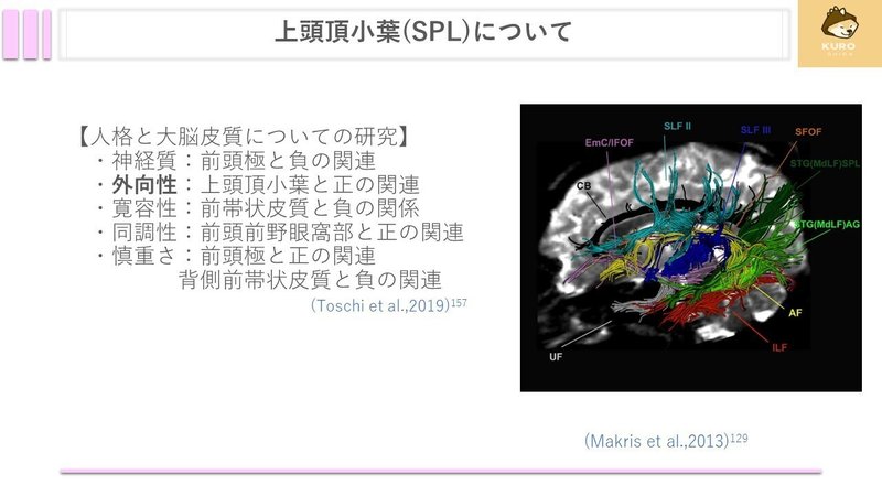
上頭頂小葉と人格との関係を調べた研究では、外向性が上頭頂小葉と正の相関があることがわかりました。
他の人格は自分の感覚であったり考えであるのに対し、外向性は
自分の内ではなく外なので、外からの様々な感覚を統合したりする上頭頂小葉が関与するようです
上頭頂小葉の損傷では感覚障害など様々な症状の他にも
頭頂葉性運動失調が挙げられます

159) 二村明徳, 河村 満 : 頭頂葉性運動失調. アクチュアル 脳・神経疾患の臨床 小脳と運動失調 : 小脳はなにをしているのか(辻 省次総編集, 西澤正豊専門編集). 中山書店, 東京, pp.289-294, 2013.
上頭頂小葉の損傷では、視覚性の入力が上頭頂小葉に届きますが、ここで障害があり、2つの型に分かれます。
Balint型が中心視野へのリーチ動作が障害され、
Garcin型が周辺視野へのリーチ動作が障害されます。
さらに上頭頂小葉から運動前野へ向けて情報が送られてリーチ動作へ繋がるので、運動前野性のリーチ動作障害にも関与しています。
対象物への脳幹の眼球運動にも関与があるようです。

161) Galletti C, Kutz DF, Gamberini M, et al. : Role of the medial parieto-occipital cortex in the control of reaching and grasping movements. Exp Brain Res, 153 : 158-170, 2003.
次は舌状回についてです。

舌状回は鳥距溝の下方部分です。
脳神経ペディアによると単語の認知もしているようです。
街並み失認や大脳色覚障害を生じることから、色や街並みを処理していると考えられます

163) Takahashi N, Kawamura M : Pure topographical disorientation─the anatomical basis of landmark agnosia. Cortex, 38 : 717-725, 2002.
164) Bartolomeo P, Bachoud-Lévi AC, Thiebaut de Schotten M. The anatomy of cerebral achromatopsia:a reappraisal and comparison of two case reports. Cortex 2014;56: 138-44.
こちらも同じような結果となっていますね

166) Epstein R, Kanwisher N. A cortical representation of the local visual environment. Nature 1998;392:598601 .
167)Epstein R. Parahippocampal and retrosplenial contributions to human spatial navigation. Trends Cogn Sci 2008; 12:388-96.
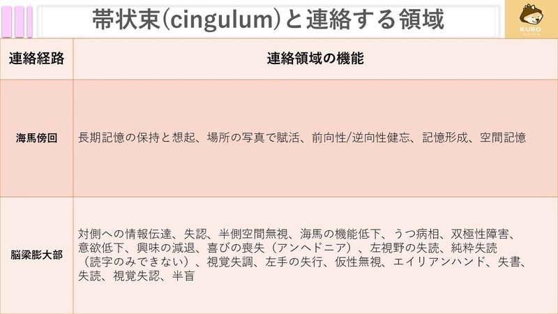
次は海馬傍回についてです。

海馬傍回の機能のまとめです。
次で詳しく解説します。

169) Epstein et al.,1998) Epstein R, Kanwisher N. A cortical representation ofthe local visual environment. Nature 1998;392:598-601
170) Epstein et al.,2008 Epstein R. Parahippocampal and retrosplenial contribu-tions to human spatial navigation. Trends Cogn Sci2008;12:388-96.
171) Reed et al.,1998 Reed JM, Squire LR. Retrograde amnesia for facts and events:findings from four new cases. J Neurosci 1998;18:3943-54.
172) Caine et al.,2000 Caine D, Watson JD. Neuropsychological and neuro-pathological sequelae of cerebral anoxia:a critical re-view. J Int Neuropsychol Soc 2000;6:86-99. 173) Eric et al., 2022) カンデル神経科学第2版 p597
174) 渡辺雅彦 2017脳神経ペディア p28
海馬傍回は長期記憶に関する部位に投射し、長期記憶に関与しています。
海馬傍回は場所の写真でも賦活されます。
また、海馬傍回を含む病巣は、前向性健忘に加え、逆向性健忘が出現し、
再認も低下します。
さらに空間の記憶にも関与するとされています。

169) Epstein et al.,1998) Epstein R, Kanwisher N. A cortical representation ofthe local visual environment. Nature 1998;392:598-601
170) Epstein et al.,2008 Epstein R. Parahippocampal and retrosplenial contribu-tions to human spatial navigation. Trends Cogn Sci2008;12:388-96.
171) Reed et al.,1998 Reed JM, Squire LR. Retrograde amnesia for facts and events:findings from four new cases. J Neurosci 1998;18:3943-54.
172) Caine et al.,2000 Caine D, Watson JD. Neuropsychological and neuro-pathological sequelae of cerebral anoxia:a critical re-view. J Int Neuropsychol Soc 2000;6:86-99. 173) Eric et al., 2022) カンデル神経科学第2版 p597
174) 渡辺雅彦 2017脳神経ペディア p28
次は脳梁膨大部です

脳梁は左右半球を結びます。
失認や半側空間無視、記憶にも関与しています。

176) (石合 純夫 (2022) 高次脳機能障害学 第3版 p124)
177) (石合 純夫 (2022) 高次脳機能障害学 第3版 p138)
178) (Eric et al., 2022 カンデル神経科学第2版 p94)
左視野の文字情報は右後頭葉から脳梁膨大部を経て左頭頂葉に入力されます。そのため脳梁膨大部損傷では言語野へ到達しないため、失読が生じます。

181) (松田 実 初学者のための神経心理学入門 2022 p121)
脳梁の離断は処理速度障害となるが記憶や実行系機能には関与せず、
運動機能と相関している。
脳梁離断症候群は多彩な高次脳機能障害が生じます。

183) Park MK, Hwang SH, Jung S, Hong SS, Kwon SB. Lesions in the splenium of the corpus callosum: Clinical and radiological implications. Neurol Asia 2014;19:79-88.
184) 大槻 美佳, 脳梁および近傍領域損傷による高次脳機能障害(<特集>側脳室腫瘍の診断と治療), 脳神経外科ジャーナル, 2009, 18 巻, 3 号, p. 179-186, 公開日 2017/06/02, Online ISSN 2187-3100, Print ISSN 0917-950X, https://doi.org/10.7887/jcns.18.179, https://www.jstage.jst.go.jp/article/jcns/18/3/18_KJ00005361635/_article/-char/ja,
最後に、帯状束についてです。

帯状束は、運動無視や記憶にも関与しています。
運動無視とは、問題がないのに運動が減少するものです。
記憶に関しては、視床前核から帯状束を通って海馬に情報を送っているため、帯状束の損傷により記憶障害も生じやすいと考えられます。

186) Fiorel l i M, Bl in J, Bakchine S, et al . PET studies of cortical diaschisis in patients with motor hemi-neglect. J Neurol Sci 1991 ; 104: 135-42.
187) Punt TD, Riddoch MJ. Motor neglect : impl ications for movement and rehabi l itation fol lowing stroke. Disabil Neurol Sci 1991 ; 104: 135-42.
188) Likitjaroen Y, Suwanwela NC, Mitchel l AJ, et al . Isolated motor neglect fol lowing infarction of the posterior l imb of the right internal capsule:a case study with diffusion tensor imaging-based tractography. J Neurol 2012;259: 100-5.
189) Migliaccio R, Bouhali F, Rastelli F, Ferrieux S, Arbizu C, Vincent S, Pradat-Diehl P, Bartolomeo P. Damage to the medial motor system in stroke patients with motor neglect. Front Hum Neurosci. 2014 Jun 11;8:408. doi: 10.3389/fnhum.2014.00408. PMID: 24966826; PMCID: PMC4052665.
190) (石合 純夫 2022 高次脳機能障害学 第3版 p147~p148)
191) (真柳佳明ら, 2018 訳 : 脳の機能解剖と画像診断, 第2版. 医学書院)
帯状束の不完全性が高いほどOCD患者の重症度が高いとされています。
また、左帯状束のFAが高いと注意機能の向上も考えられます。

193) Sweet JA, Gao K, Chen Z, Tatsuoka C, Calabrese JR, Sajatovic M, Miller JP, McIntyre CC. Cingulum bundle connectivity in treatment-refractory compared to treatment-responsive patients with bipolar disorder and healthy controls: a tractography and surgical targeting analysis. J Neurosurg. 2022 Jan 21:1-13. doi: 10.3171/2021.11.JNS211833. Epub ahead of print. PMID: 35061996; PMCID: PMC10193487.
196) Archer DB, Moore EE, Pamidimukkala U, Shashikumar N, Pechman KR, Blennow K, Zetterberg H, Landman BA, Hohman TJ, Jefferson AL, Gifford KA. The relationship between white matter microstructure and self-perceived cognitive decline. Neuroimage Clin. 2021;32:102794. doi: 10.1016/j.nicl.2021.102794. Epub 2021 Aug 28. PMID: 34479171; PMCID: PMC8414539.
幻覚がある統合失調症患者では左帯状束の不完全性が高いようです。
また、加齢によってもFAが低下すると考えられます。
また、2型糖尿病患者、ASDで帯状束の完全性の低下がみられます。

192) Sibilia F, Kehoe EG, Farrell D, et al. Aging-Related Microstructural Alterations Along the Length of the Cingulum Bundle. Brain Connect. 2017;7(6):366-372. doi:10.1089/brain.2017.0493
193) Sweet JA, Gao K, Chen Z, Tatsuoka C, Calabrese JR, Sajatovic M, Miller JP, McIntyre CC. Cingulum bundle connectivity in treatment-refractory compared to treatment-responsive patients with bipolar disorder and healthy controls: a tractography and surgical targeting analysis. J Neurosurg. 2022 Jan 21:1-13. doi: 10.3171/2021.11.JNS211833. Epub ahead of print. PMID: 35061996; PMCID: PMC10193487.
198) Mårtensson J, Lätt J, Åhs F, et al. Diffusion tensor imaging and tractography of the white matter in normal aging: The rate-of-change differs between segments within tracts. Magn Reson Imaging. 2018;45:113-119. doi:10.1016/j.mri.2017.03.007
199) Cui Y, Tang TY, Lu CQ, Cai Y, Lu T, Wang YC, Teng GJ, Ju S. Abnormal Cingulum Bundle Induced by Type 2 Diabetes Mellitus: A Diffusion Tensor Tractography Study. Front Aging Neurosci. 2020 Dec 11;12:594198. doi: 10.3389/fnagi.2020.594198. PMID: 33384593; PMCID: PMC7771529.
200) Ameis SH, Fan J, Rockel C, Soorya L, Wang AT, Anagnostou E. Altered cingulum bundle microstructure in autism spectrum disorder. Acta Neuropsychiatr. 2013;25(5):275-282. doi:10.1017/neu.2013.2
201) Ikuta T, Shafritz KM, Bregman J, Peters BD, Gruner P, Malhotra AK, Szeszko PR. Abnormal cingulum bundle development in autism: a probabilistic tractography study. Psychiatry Res. 2014 Jan 30;221(1):63-8. doi: 10.1016/j.pscychresns.2013.08.002. Epub 2013 Nov 11. PMID: 24231056; PMCID: PMC3918471.
202) Ezzati A, Katz MJ, Lipton ML, Zimmerman ME, Lipton RB. Hippocampal volume and cingulum bundle fractional anisotropy are independently associated with verbal memory in older adults. Brain Imaging Behav. 2016 Sep;10(3):652-9. doi: 10.1007/s11682-015-9452-y. PMID: 26424564; PMCID: PMC4816657.
パーキンソン病でも帯状束の完全性の低下がみられ、認知機能・精神機能に関与しています。

204) Oestreich LKL, Wright P, O'Sullivan MJ. Microstructural changes in the reward system are associated with post-stroke depression. Neuroimage Clin. 2020;28:102360. doi: 10.1016/j.nicl.2020.102360. Epub 2020 Jul 22. PMID: 32795963; PMCID: PMC7426585.
205) Migliaccio R, Bouhali F, Rastelli F, Ferrieux S, Arbizu C, Vincent S, Pradat-Diehl P, Bartolomeo P. Damage to the medial motor system in stroke patients with motor neglect. Front Hum Neurosci. 2014 Jun 11;8:408. doi: 10.3389/fnhum.2014.00408. PMID: 24966826; PMCID: PMC4052665.
帯状束について、論文を集めてみました。
いかがだったでしょうか。
また連合線維を追っていくので気になる方はチェックしてみてください。
ご覧になっていただきありがとございました!
この記事が気に入ったらサポートをしてみませんか?