WGA:White Globe Appearance

内視鏡読影:NBI拡大画像 胃がん WGAについて
Doyama, H., N. et al. "The "White Globe Appearance" (Wga): A Novel Marker for a Correct Diagnosis of Early Gastric Cancer by Magnifying Endoscopy with Narrow-Band Imaging (M-Nbi)."
Endosc Int Open 3, no. 2 (Apr 2015)より転載・引用
今回は土山先生の執筆された論文から読影していきたい。
【読影】
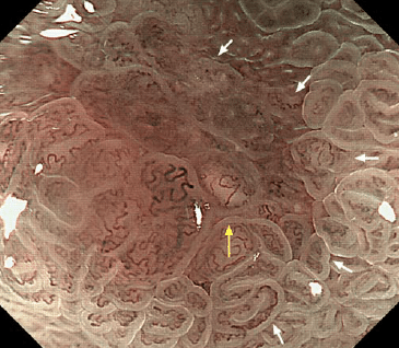
NBI拡大観察の写真である。
白矢印の内側の胃腺窩構造は不明瞭であり、表層血管の拡張と蛇行を認める。
demarcation line(癌と非癌部境界)は認識できており、正常病変と腺窩構造が消失している部位の領域は明瞭であり、これは組織型が異なる部位が存在していることを示している。
白矢印の外側には上皮表層に青白い光の線(light blue crest)と窩間部にやや白色沈着物(white opaque substance)を認めていることから、腸上皮化生が背景粘膜にある考えられる。
この画像だけではピロリ菌感染中または除菌後粘膜かの鑑別は不可能だが、腸上皮化生が認めることから、以前にピロリ菌感染による慢性炎症が持続していたことは間違いないだろう。
内視鏡読影:NBI
(説明文)
Representative endoscopic image of the white globe appearance
(WGA) visualized by magnifying endoscopy with narrow-band imaging
(M-NBI) with maximal magnification.
The WGA inside cancerous mucosa(yellow arrow) was located close to the demarcation line between the cancerousmucosa and the surrounding mucosa (white arrows), indicative of“marginal distribution.” The WGA features were a whitish color less intenseat the lesion’s periphery than in the center (reflecting its globular shape),and the presence of overlying microvessels. The WGA was approximately0.5mmin size. The lesion size was determined by observation using a visualfield of approximately 3.4mm with maximal magnification as a guide, withthe black hood mounted on the endoscope.
ndosc Int Open 3, no. 2 (Apr 2015)より転載・引用
内視鏡診断で一発診断という項目があり、その一つにWGA(White Globe Appearance)がある。
これは言葉の通り、白色の球状物体である。
WGAの周囲(白矢印の示す範囲内)は不整構造があり、それらから胃がんであると診断できる。
WGA:特異度が高く(約98%)、これがあればほぼ胃癌と診断できる項目となる。※特異度が高いということは、これがあれば確定診断できる。
【病理写真】
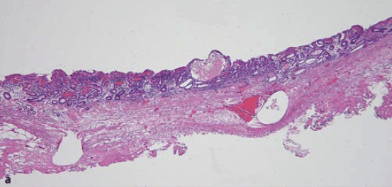
Doyama, H., N. et al. "The "White Globe Appearance" (Wga): A Novel Marker for a Correct Diagnosis of Early Gastric Cancer by Magnifying Endoscopy with Narrow-Band Imaging (M-Nbi)." Endosc Int Open 3, no. 2 (Apr 2015)より転載・引用
この記事が気に入ったらサポートをしてみませんか?